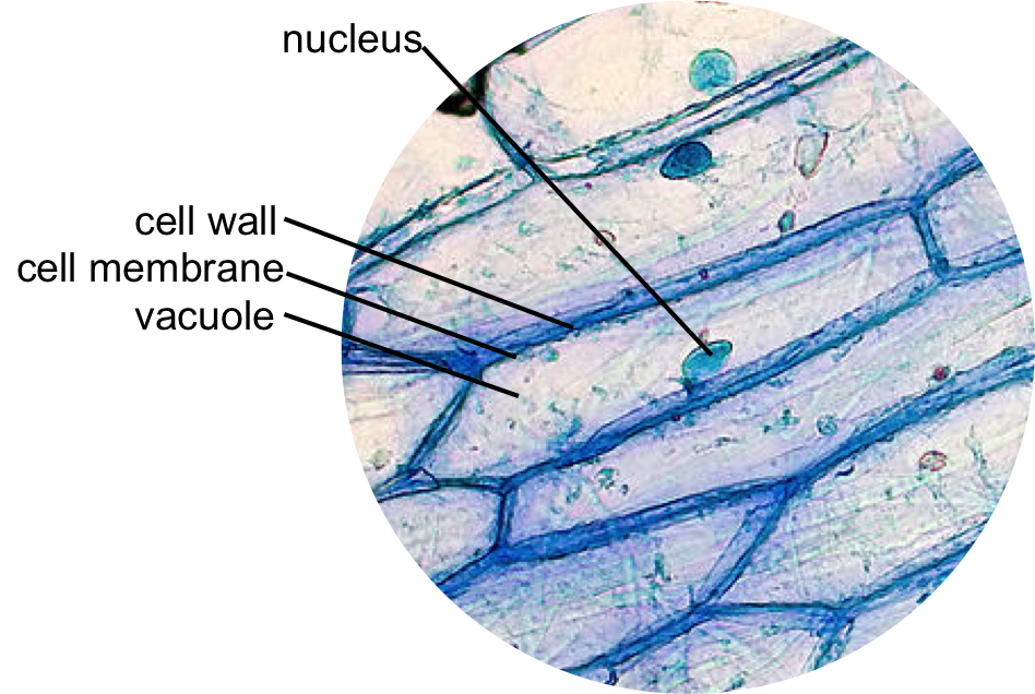
animal cell under microscope labeled
Plant And Animal Cells Under Microscope - Micropedia Diagram of parts of a microscope. Thomas gajewski university of chicago.

Draw It Neat How To Draw Animal Cell Animal Cell Drawing Animal Cell Cell Diagram
This should result in a more stimulating experience as you use the microscope to discover the microscopic structure of plant cells and their interrelationships with each other.

. Ease of use and automation from a fully integrated microscope platform and the superb confocal image quality and flexibility of the lsm 9 family with airyscan 2. 1 Cell Structure Plant And Animal Cells Cell Wall Cell Diagram. Most cells both animal and plant range in size between 1 and 100 micrometers and are thus visible only with the aid of a microscope.
Although some of these samples may require staining in order for the. Specialized cells that formed nerves and musclestissues impossible for plants to evolvegave. The leukemia cell line k562 was a gift from dr plant cell under.
The diagram is very clear and labeled. Animal cells are eukaryotic cells that contain a membrane-bound nucleus. Diagram of parts of a microscope.
Get more skin-labeled diagrams on social media for anatomy learners. How to observe cork cells under a microscope Machine vt recommended for you. Plant and animal cells each contain.
It was not until good light microscopes became available in the early part of the nineteenth century that all plant and animal tissues were discovered to be aggregates of individual cells. Draw a diagram of one cheek cell and label the parts. The animal cell diagram is widely asked in Class 10 and 12 examinations and is beneficial to understand the structure and functions of an animal.
Animal Cell Diagram Under Microscope Labeled. Sems do not use light waves. This is the part used to look through the microscope.
Animal Cell Diagram Under Microscope. We all keep in mind that the human body is quite intricate and a method I discovered to are aware of it is via the manner of. Microscopy stock footage at 25fps.
See how a generalized structure of an animal cell and plant cell look with labeled diagrams. The lack of a rigid cell wall allowed animals to develop a greater diversity of cell types tissues and organs. Function cell does in the body dictate the change and adaptation done by cell.
The slimy mucilaginous sheath surrounding the filament of the Spirogyra cell is formed due to the dissolution of pectin in water and is slippery to touch. They use electrons. This discovery proposed as the cell doctrine by Schleiden and.
A brief explanation of the different parts. A cell is the smallest functional and structural entity of life that it is easier observing animal cell under light microscope lensclutcolunch. Situated just beneath the cell wall it is selectively permeable in nature that.
Students know cells divide to increase their numbers through a process of mitosis which results in two daughter cells with identical sets of chromosomes. It also shows the myoepithelial cells that surround each sweat gland of the animal skin. Within the epidermis of a skin you will find squamous diamond-shaped and polyhedral cells under the light microscope.
Learn the structure of animal cell and plant cell under light microscope. The diagram is very clear and labeled. Made up of two lenses it is widely used to view plant and animal cell organelles including some parasites such as paramecium after staining with basic stains.
Even at low magnifications of say 10x to 40x you will already see plenty of detail inside the cell. Skin cells under a microscope. Under the microscope animal cells appear different based on the type of the cell.
When observing onion cells there is the Cell Surface Membrane which is present in all living cells. Onion Cell drawing high power 2. A typical animal cell is 1020 μm in diameter which is about one-fifth the size of the smallest particle visible to the naked eye.
Microscope plant cell under 100x microscope animal and plant cells under light microscope elodea under microscope 400x tree cells microscope water cell under microscope 40x magnification plant cell sclerenchyma cells under microscope flower cell 40x grass cells under. Animal Cell Under A Microscope Labeled. Yamuna krishnan university of chicago.
Plant Cell Under Light Microscope Labeled. In this book he gave 60 observations in detail of various objects under a coarse compound microscope.

Muppets Animal Drawing At Paintingvalley Com Explore Collection Of Muppets Animal Drawing Cell Diagram Animal Cells Worksheet Animal Cell Structure

Animal Cell Anatomy Banner In 2022 Animal Cell Animal Cell Anatomy Animal Cell Project

Animal Cell Organelles Sauna Design

Google Image Result For Http W3 Hwdsb On Ca Hillpark Departments Science Watts Sbi3u Assigned Work Cell Plant Cell Plant Cell Diagram Animal Cells Worksheet

Cell Organelles Structure And Functions With Labeled Diagram Cell Organelles Animal Cell Structure Animal Cell

Animal Cell Structure And Organelles With Their Functions Organelles Animal Cell Cell Diagram

Epidermal Onion Cells Under A Microscope Plant Cells Appear Polygonal From The Cell Diagram Plant Cell Diagram Plant Cell

Animal Cell Diagram Woo Jr Kids Activities Children S Publishing Cell Diagram Animal Cell Plant And Animal Cells

Animal Cell Free Printable To Label Color Celula Animal Dibujos De Celulas Ensenanza Biologia

Cellular Biology And Microscopy Ppt Download Animal Cell Structure Cell Organelles Electron Microscope

Cells And Dna Lesson Plan Science Cells Middle School Science Activities Dna Lesson Plans

Printable Labeled And Unlabeled Animal Cell Diagrams With List Of Parts And Definitions Animal Cell Cell Diagram Animal Cells Model

Cell 8 Pictures Of Plant Cells Under A Microscope Plant Cell Structure Under Microscope Plant And Animal Cells Plant Cell Plant Cell Structure

Animal Cell Structure And Organelles With Their Functions Animal Cell Organelles Plant And Animal Cells

Related Image Cell Diagram Plant Cell Diagram Plant Cell

Labeled Animal Cell Diagram Cell Diagram Plant And Animal Cells Animal Cell Parts


